


Welcome / Velkommen
Our research focusses on basic molecular mechanisms and new treatment options for neurodegenerative diseases with special emphasis on Alzheimer’s disease. We investigate clearance mechanisms of toxic peptides via the blood-brain barrier, new imaging techniques, mitochondrial function, and neuroimmunological mechanisms. Our work also includes Parkinson’s, Huntington’s, ALS and other proteopathies of the brain. We also investigate brain tumors and brain metastasis using Mass Spectrometry imaging. If you are interested to collaborate with us, please do not hesitate to get in contact.
News

Advanced visualizations and new molecular maps reveal the brain’s hidden chemisty.
Informasjoner på Norsk på OUS og UiO sine nettsider.
Read Science Advances 12(35) article – 26. Aug 2026.
We have developed new ways to see how thousands of molecules are distributed in the brain or other organs. These maps could improve our understanding of dementia, cancer and other diseases and may eventually support earlier and more precise diagnosis and patient-oriented treatment development.
A chemical map of tissue
Conventional microscopy shows the shape of cells and tissue. The new approach – mass spectrometry imaging (MSI) – also shows where different molecules are located. Each point in a tissue sample receives a chemical fingerprint, and the points are combined to form a detailed map. We have created digital tools that make these huge and complex datasets easier to understand through colorful images. We can compare different parts of the tissue, find small disease-related changes and investigate which molecules characterise specific areas. Our new mouse lipid atlas maps the chemical organisation of >100 brain regions in the mouse brain to help understanding function and organization.
The aim is to see signs of disease before they become visible in conventional tissue images.
Why lipids matter
Lipids are essential building blocks in the brain but also in other organs and cancer. They form part of cell membranes and help cells communicate. Several known risk factors for Alzheimer’s disease affect how lipids are transported and handled. The atlas shows that lipids follow precise patterns. Brain regions that work together often have very similar chemical features. Researchers can now study the chemistry around deposits associated with Alzheimer’s disease or other dementias. This will offer new explanations for why some parts of the brain are more vulnerable than others and are effected earlier or more severe.


Two sagittal mouse brain MSI data visualization examples of the same dataset (res. 30µm/pixel).
Potential across several diseases
The new methods can also be used to investigate brain tumours, other cancers, metastasis, inflammation, and kidney disease. A molecular map can show differences between diseased and healthy tissue, between different parts of a tumour and between areas that respond differently to treatment. The technology is still a research tool and must be tested thoroughly on patient samples before it can be used in routine diagnosis. The aim is not to replace the microscope, but to give doctors and pathologists an additional layer of information about what is happening inside tissue.
This will improve diagnostics but also patient-oriented therapy. Through truthful visualizations of MSI data, we enable determination of cancer subentities with specific metabolic pattern. These methods can also be used to diagnose rare childhodd diseases, such as metabolic muscle diseases.

Visualization example of a MSI human kidney biopsy dataset (res. 5µm/pixel).
A new view of disease
Where we previously saw mainly what tissue looked like, we can now begin to see the hidden chemistry und function behind health, aging and disease. In the longer term, this could support earlier diagnosis, better treatment choices and more
precise monitoring of patients.
Read Science Advances article about truthful visualizations of high dimensional MSI data.
Read Free Neuropathology article about the establishment of a brain lipid atlas.
Read BioaRxiv preprint with enhanced methods and automated pathology panels.
Listen to our PodCasts.
Email us.
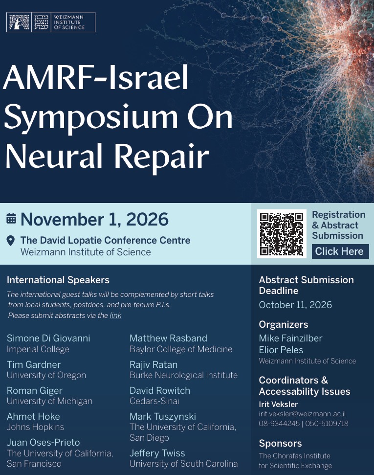
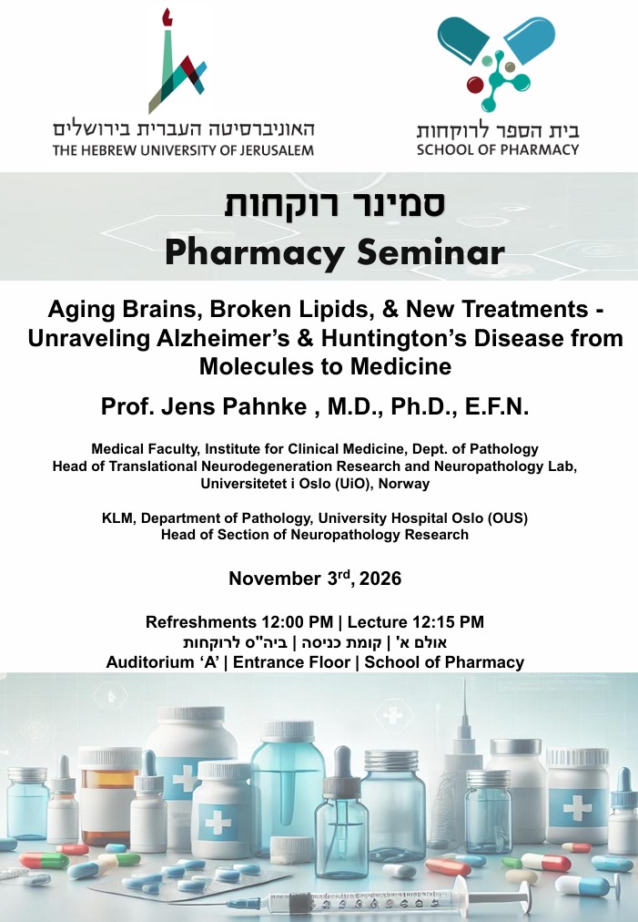
Announcement posters:


Further talks about aging, dementia, treatment, and new mass spectrometry methods:
01.11.2026 Rehovot (AMRF26 meeting on Neurorepair (13:00) – see above)
03.11.2026 Jerusalem (Seminar (12:00) at Hadassah Medical Center – see above)
04.11.2026 Rehovot (Seminar (14:30) at Weizmann Inst., Dept. of Mol. Neurosci. – see above)
05.11.2026 Tel Aviv (Plenary talk (13:35) at Israeli Neuroimmunology Society congress – Y. Rabin Center – pdf)
*
10.01.2027 Eilat (Talk at INSF27 congress)
14.01.2027 Rehovot (Seminar at Weizmann Inst., Dept. of Chemisty)
*
11.03.2027 Frankfurt (Talk at EPHAR27 Congress)
recent:
31.09.2026 Oslo (No-Age/AD congress)
07.09.2026 Sapporo (Hokkaido University research seminar – see below)
09.09.2026 Sapporo (ICATS-FHM 2026 congress)

Johanniskraut und Griechischer Bergtee zur Demenzbehandlung
Hier ein Link zu Rückmeldungen von Patienten und Angehörigen zu Behandlungen bei dementieller Symptomatik absierend auf unseren Experimenten mit Alzheimer-Mäusen. Publizierte wissenschaftliche Daten finden Sie unter Publikationen.
Externe Info-Seite zu Griechischem Bergtee
St. John’s wort and Greek Mountain tea for dementia treatment
Here you find a link to replies of patients and relatives to treatment that we have developed for dementia based on our Alzheimer mice experiments. Published scientific articles you can find under publications.
Information for patients and their relatives
How patients can help us:
Our research needs research funding – if you like what we do, please support patient initiatives or scientific organisations which support research matters.
Or donate to us directly as a free donation or bound to a specific purpose: dementia, Alzheimer’s, brain tumors, Huntngton’s or even more specific.
We will drive your funded and guided projects. Check our funding page for ongoing and recent projects.
Many thanks for supporting dementia research.
Please contact us for questions:
Email: info@pahnkelab.eu