<< back
Clinical and research pathology use microscopy as a basic tool for diagnosis and/or evaluation of disease progression or treatment. However, this approach requires quantification or cell classification, which often entails counting thousands of cells/objects. Thus, image analysis in this field is tedious and demands a great amount of personnel and time investment. In addition, the traditional approach implies a bias as classification of cells/objects depends only on the pathologist’s training. Nowadays, advancements in computer engineering have enabled computer training known as artificial intelligence (AI) and machine learning. Thus, computers can be trained to perform some tasks based on a training provided previously. Machine learning in pathology can reduce the time and personnel invested in image analysis. At the same time, analysis of pathology images by AI applies the same criteria to all images independently of previous patient/sample information, can overcome image artefacts and does not show signs of fatigue after hours of analysis. Thus, AI delivers unbiased reports in a reduced time compared to the traditional approach.
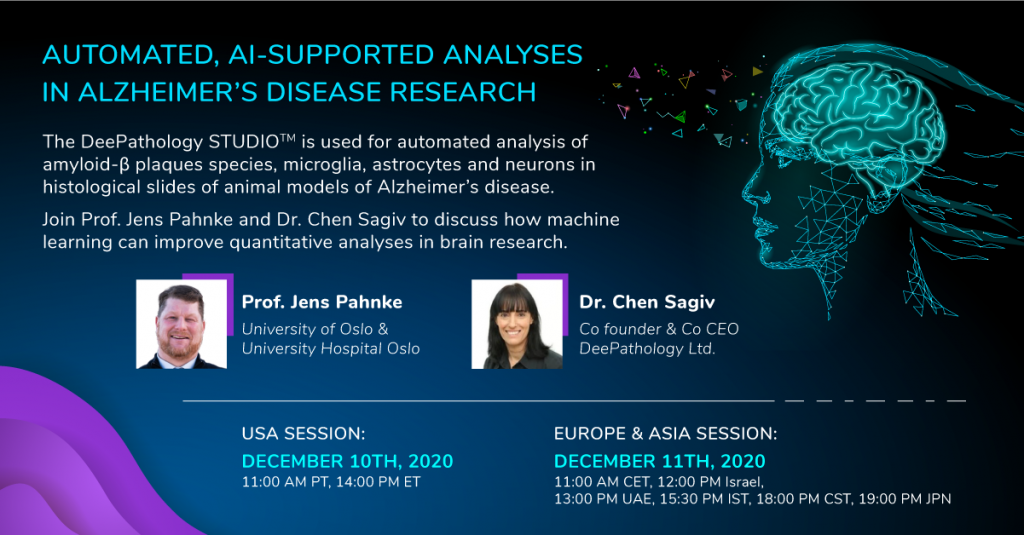
Currently, our group develops tools to automatically assess morphological changes in histological sections for diagnostic and research purposes together with DeePathology.AI Ltd.. This approach has shown a 90% reduction of time invested for analysis.
* Published Research Papers (selection):
-
Development of deep learning models for microglia analyses in brain tissue using DeePathology™ STUDIO
Journal of Neuroscience Methods (2021)
Möhle L, Bascuñana P, Brackhan M, Pahnke J
364:109371 PubMed PDF
-
Machine Learning – supported analyses improve quantitative histological assessments of β-amyloid deposits and activated microglia
Journal of Alzheimer’s Disease (2020)
Bascuñana P, Brackhan M, Pahnke J
79(2):597-605. PubMed PDF
